You went in because your knee was hurting. You came out with three words you can't stop thinking about.
“Bone on bone.”
Maybe a GP said it. Maybe the radiographer pointed at the X-ray and used the phrase. Maybe a friend who saw your scan said it out loud. However it landed, the words sat in your head on the drive home and they probably haven't fully left since.
You started doing the maths. How long until you can't take the stairs. How long until you can't walk the dog. Whether you'll be one of the people who ends up with a replacement, and how soon. Whether to stop the things you love now, in case you're making it worse.
Here's what we want you to know before you read another paragraph. The phrase you heard is real, but it doesn't mean what you almost certainly think it means. And the path forward — the one that the Australian clinical guidelines actually recommend you try first — is far less dramatic, far less invasive, and far more effective than most patients are ever told.
What “bone on bone” actually means on an X-ray
When someone says your knee is “bone on bone,” what they actually mean is that on the X-ray, the visible gap between your thigh bone (the femur) and your shin bone (the tibia) has narrowed. That gap, in a healthy knee, is occupied by cartilage. When the cartilage thins, the gap closes. Closer bones on the X-ray, less cartilage in the joint.
What it does not mean is that your bones are literally grinding against each other every step you take. It does not mean your knee is “destroyed.” It does not mean you have run out of options. It's a poor predictor of how much pain you actually feel, and an even poorer predictor of how well you can move.
Why what you saw on the X-ray isn't the whole story
A systematic review of imaging and pain in knee osteoarthritis, summarising the literature across multiple studies, found a weak and inconsistent relationship between what the X-ray shows and what the patient experiences.2 Some people with the worst-looking X-rays walk three kilometres a day with minimal symptoms. Other people whose X-rays look mild can barely climb a flight of stairs.3
Australia's 2024 Osteoarthritis of the Knee Clinical Care Standard puts this in clinical guidance for every GP in the country. The standard explicitly states that imaging should not be used as the primary tool to diagnose or grade knee osteoarthritis, because doing so leads to overtreatment of structural findings that aren't clinically meaningful.1 Translated, the people who set the rules for treating knee arthritis in Australia have told doctors to stop putting so much weight on the X-ray.
So when you stare at the image and the radiology report and feel your future shrinking, please understand: the picture you are looking at is not the variable that decides how you feel, what you can do, or what you can still get back. The variables that decide those things are the ones we're about to talk about. And every one of them is changeable.
What is actually driving your knee pain
A knee is not just cartilage. A knee is a joint surrounded by muscle, supported by ligaments and tendons, regulated by a synovial lining, and loaded by the way you stand and walk and sit. Pain in an arthritic knee can come from any of these structures, and most of them are not what the X-ray was showing.
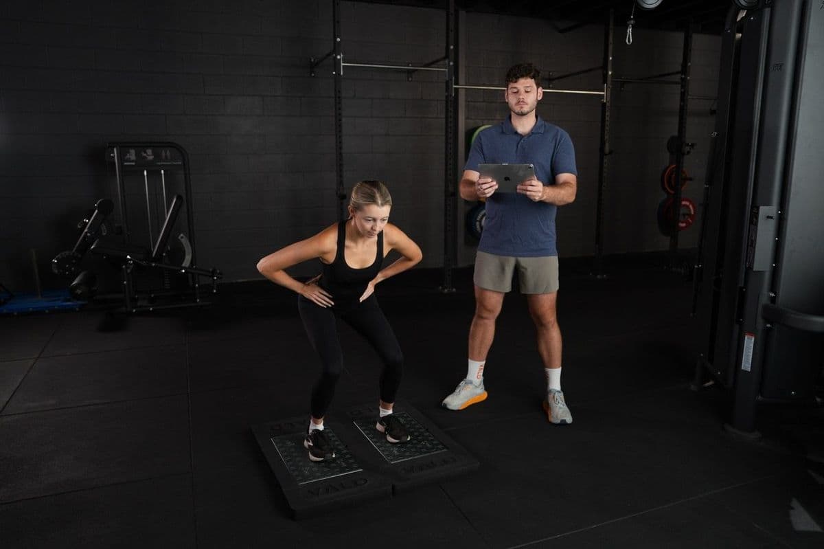
The two biggest drivers of pain and disability in knee osteoarthritis are not the cartilage status itself. They are quadriceps weakness and inflammation. Both are highly modifiable.
Your quadriceps muscles — the big muscles on the front of your thigh — are the primary shock absorbers of your knee. When they work properly, they take load off the joint surface every time you take a step, climb stairs, or stand up from a chair. When they weaken, the joint surface absorbs more of the load itself. Weak quads make a sensitive knee more sensitive. Strong quads make a sensitive knee feel dramatically better, often within weeks.10
Inflammation is the other half of the picture. Osteoarthritis is no longer thought of as a purely mechanical “wearing out” process. It's now understood as the joint working harder to repair and regulate itself, with periodic inflammation contributing to flare ups of pain, stiffness and swelling. Inflammation responds to load, to movement, to general health, and to consistent activity. It also responds to giving the joint a break from sudden, unfamiliar stress. Both ends of that are within your control.